Blog Livia

Welcome to My Blog! 만나서 반갑습니다 :)
Showing posts with label Salivary Gland Diseases- Reactive Lesions. Show all posts

Monday, November 1, 2010

Maxillary Sinus Retention Cyst/ Pseudocyst

Retention cysts and pseudocysts involving the lining of the maxillary antrum are common findings in panoramic radiographs. These lesions are discovered incidentally and are of little clinical significance.

Etiology and Pathogenesis

Retention cysts are thought to arise from blockage of an antral seromucous gland, resulting in a ductal epithelium-lined cystic structure filled with mucin. Pseudocysts are inflammatory in origin and result from fluid accumulation within the sinus membrane. They may be related to infection or allergy. Bacterial toxins, anoxia, or other factors presumably cause leakage of protein into surrounding soft tissue, thus raising the extravascular osmotic pressure with subsequent fluid increase. 

Clinical Features 
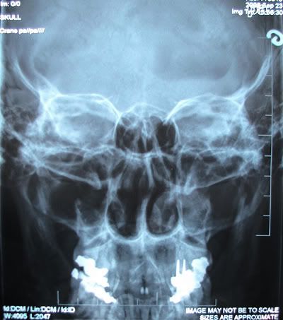
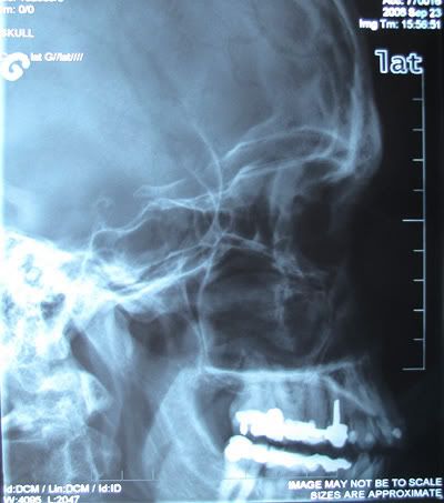
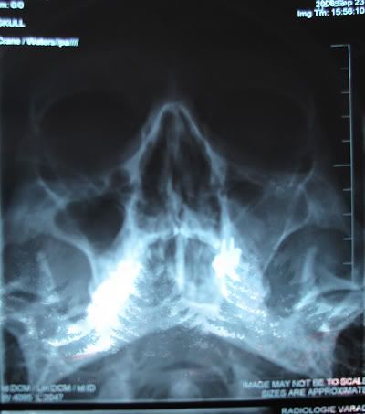
The great majority of these lesions are asymptomatic, although there maybe some slight tenderness in the mucobuccal fold or, more rarely, palpable buccal expansion in this region. In panoramic and periapical radiographs, retention cysts and pseudocysts of the maxillary sinus are hemispheric, homogeneously opaque, and well delineated. They usually demonstrate an attachment to the floor of the antrum, with size being a function of the anatomic space rather than of duration, Uncommonly these lesions may appear bilaterally.


Histopathology

The pathogenesis of the two forms of antral cysts is reflected in the histologic appearance. The retention cyst is lined by pseudostratified columnar epithelium with occasional mucous cells interspersed. The supportive elements are minimally inflamed. The pseudocyst shows no evidence of an epithelial lining but, rather, pools of mucoid material surrounded by slightly compressed connective tissue.


Differential Diagnosis
A clinical differential diagnosis of cysts and pseudocysts arising within the mucosa of the maxillary sinus would include polyps, hyperplasia of the sinus lining secondary to odontogenic infection, maxillary sinusitis, and neoplasms arising within the soft tissues of the antral lining.

Treatment
 Antral retention cysts and pseudocysts are generally left untreated because they are limited in growth and are not destructive, and most spontaneously rupture. Therefore periodic observation is all that is required.

Mucus Retention Cyst

Etiology and Pathogenesis
Mucus retention cysts result from obstruction of salivary flow because of a sialolith, periductal scar, or impinging tumor. The retained mucin is surrounded by ductal epithelium, giving the lesion a cystlike appearance microscopically. Sialolithiasis. Obstruction due to a salivary stone, or sialolith, is usually associated with the submandibular gland. The sialolith(s) may be found anywhere in the ductal system from the gland parenchyma to the excretory duct orifice. A sialolith represents the precipitation of calcium salts (predominantly calcium carbonate and calcium phosphate) around a central nidus of cellular debris or inspissated mucin. 

Clinical Features
Mucus retention cyst is less common than mucus extravasational phenomenon. It usually appears in an older age-group and is most commonly seen in the upper lip, palate, cheek, and floor of the mouth. Lesions present as asymptomatic swellings, usually without antecedent trauma. They vary in size from 3 to 10 mm and on palpation are mobile and nontender. The overlying mucosa is intact and of normal color. Mucin in floorof- mouth lesions may penetrate musculature and escape in to the soft tissues of the neck, causing a "plunging ranula."


Histopathology
Histopathology. The cystlike cavity of a mucus retention cyst is lined by normal but compressed ductal epithelial cells. The type of lining formed by the epithelial cells ranges from pseudostratified to stratified squamous. The cyst lumen contains inspissated mucin or a calcified sialolith. The connective tissue around the lesion is minimally inflamed, although the associated gland shows obstructive change.

Differential Diagnosis
Salivary gland neoplasms, mucus extravasation phenomenon, and benign connective tissue neoplasms should be included in a clinical differential diagnosis. Dermoid cyst might also be included for lesions in the floor of the mouth.

Treatment and Prognosis
Treatment requires removal of the mucus retention cyst and the associated minor salivary gland to avoid postoperative mucus extravasation phenomenon. Lesions of the major salivary glands can be treated in a similar way or, on occasion, only by removal of the obstruction (sialolith) if it occurs in the distal part of the ductal system. The sialolith is either surgically removed or milked through the duct orifice. If a duct is surgically entered, special precautions are used to aid the healing process so that duct scarring is minimized. Constriction of the duct through excessive scar formation could result in recurrence. Marsupialization by the placement of a silk suture in the roof of a large mucus retention cyst, particularly in one arising in the floor of the mouth, can be useful to reduce its size before surgical excision.

Sunday, October 31, 2010

Salivary Gland Diseases- Reactive Lesions

Reactive Lesions
Mucocele is a clinical term that includes mucus extravasation phenomenon and mucus retention cyst. Because each has a distinctive pathogenesis and microscopy, they are considered separately. Ranula is a clinical term that also includes mucus extravasation phenomenon and mucus retention cyst, but it occurs specifically in the floor of the mouth. Ranula is associated with the sublingual or submandibular glands and presents as a fluctuant, unilateral,soft tissue mass. It often exhibits a bluish color that has been compared to a frog's belly; hence the term ranula. When it is significantly large, it can produce medial and superior deviation of the tongue. It may also cross the midline if the retained mucin dissects through the submucosa. A deep, so-called plunging ranula develops if mucus herniates through the mylohyoid muscle and along the fascial planes of the neck. On rare occasions it may progress into the mediastinum.

The Salivary glands Disease which are included into Reactive Lesions:
1. Mucus Extravasation phenomenon/ Mucocele
2. Mucus Retention cyst/ Ranula
3. Maxillary Sinus Retention Cyst/ Pseudocyst
4. Necrotizing Sialometaplasia
5. Adenomatoid Hyperplasia 

1. Mucus Extravasation Phenomenon
Etiology and Pathogenesis
The cause of mucocele is traumatic severance of a salivary gland excretory duct, resulting in mucus escape, or extravasation, into the surrounding connective tissue. An inflammatory reaction of neutrophils followed by macrophages ensues. Granulation tissue forms a wall around the mucin pool, and the contributing salivary gland undergoes inflammatory change. Ultimately, scarring occurs in and around the
gland.

Clinical Features
The lower lip is the most common site of mucocele, but the buccal mucosa, anterior-ventral surface of the tongue (location of Blandin-Nuhn glands), floor of the mouth, and retromolar region are often affected. Lesions are uncommonly found in other intraoral regions where salivary glands are located, probably because of a lower susceptibility to trauma. Mucus extravasation phenomenon presents as a relatively painless smooth-surfaced mass ranging in size from a few millimeters to 2 cm in diameter. It has a bluish color when mucin is superficially located. Adolescents and children are more commonly affected than adults. Lesions may fluctuate in size because of mucosal rupture over the pooled mucin. Continued production of mucin leads to recurrence. The maximum size is usually reached within several days after injury, and a viscous material is found if aspiration is attempted. Superficial mucocele is a variant of the extravasation type mucocele. Rather than arising from traumatic duct rupture, this form of mucocele is believed to arise as a result of increased pressure in the outer most part of the excretory duct. These lesions are asymptomatic and numerous, occurring most commonly in the retromolar area, soft palate, and posterior buccal mucosa. Their clinical appearance suggest a vesiculobullous disease, but the lesions persist for an extended time. Other than being a diagnostic challenge, they are of little significance.

                                                      retention mucocele on the lower lip


Histopathology
Extravasation of free mucin incites an inflammatory response that is followed by connective tissue repair. Neutrophils and macrophages are seer and granulation tissue forms around the mucin pool. The adjacent salivary gland whose duct was transected shows ductal dilation, chronic inflammation, acinar degeneration, and interstitial fibrosis.
                  histopathologic image of extravasation type mucocele from lower lip


Differential Diagnosis 
Although a history of a traumatic event followed by development of a bluish translucency of the lower lip is characteristic of mucus extravasation phenomenon, other lesions might be considered when a typical history is absent. These include salivary gland neoplasm (especially mucoepidermoid carcinoma), vascular malformation, venous varix, and soft tissue neoplasm such as neurofibroma or lipoma. Rarely, a mucocele may appear in the alveolar gingival mucosa. When this is the case, an eruption cyst or gingival cyst should be included in the differential diagnosis.

Treatment and Prognosis
The treatment of mucus extravasation phenomenon is surgical excision. Aspiration of the fluid content provides no lasting clinical benefit. Removal of the associated minor salivary glands along with the pooled mucus is necessary to prevent recurrence. No treatment is required for superficial mucoceles, since they rupture spontaneously and are short-lived.
                                                      Surgical excision of the mucocele